
LUNG CANCER AND CHEMISTRY BRANDS
Cancer is the world’s second‑leading cause of death, responsible for approximately 16.7% of global mortality (WorldPopulationClock 2026; WHO Global Health Estimates). In our Health & Chemistry Brands category, we analyze modern illnesses through chemical mechanisms, APIs, brand evolution, and real‑world sentiment.
Lung cancer treatment has evolved dramatically over the past decades. Before the rise of targeted therapies and immunotherapy, chemotherapy and radiation were the primary options — foundational treatments that remain essential today for many patients and are still widely used in combination with modern drugs. This page explores the brand‑level competition among today’s targeted therapies, tracking their popularity, social media presence, and real‑world patient experiences to reveal the rise and fall of the most influential APIs in lung cancer.
Understanding these chemical pathways helps explain why cancer forms, how treatments work, and how public interest in cancer medications rises and falls across Google Trends, Reddit, and Twitter (X).
1. Chemistry Behind Lung Cancer
Lung cancer begins with chemical damage to DNA, often caused by harmful chemical agents found in tobacco smoke, air pollution, industrial environments, and certain occupational exposures. Many of these substances are formally classified as known human carcinogens by the American Cancer Society and the International Agency for Research on Cancer (IARC).
Authoritative Carcinogen Lists
- ACS – Known and Probable Human Carcinogens https://www.cancer.org/cancer/risk-prevention/understanding-cancer-risk/known-and-probable-human-carcinogens.html#lists-of-known-human-carcinogens
- IARC Monographs – Carcinogen Classifications https://monographs.iarc.who.int/list-of-classifications
These lists include several chemical agents strongly associated with lung cancer, such as:
- Benzo[a]pyrene (polycyclic aromatic hydrocarbon)
- NNK and NNN (tobacco‑specific nitrosamines)
- Arsenic
- Asbestos fibers
- Silica dust
- Diesel exhaust particles
- Radon decay products
These substances damage DNA through oxidation, adduct formation, or chronic inflammation.
Nicotine → NNK → DNA Adduct Pathway (Chemical Mechanism)
Nicotine itself is not the primary carcinogen, but it undergoes metabolic transformations that produce highly reactive nitrosamines. See the graphic.
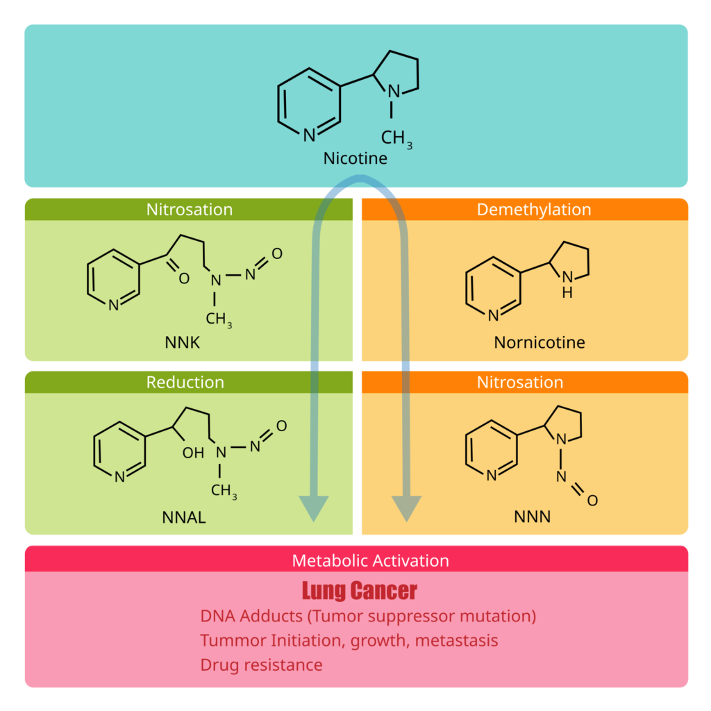
nicotine and nornicotine form NNK and NNN, two of the most potent lung carcinogens known.
- NNK is further metabolized into NNAL, which can persist in lung tissue for weeks.
- These nitrosamines undergo metabolic activation, forming electrophilic intermediates that bind covalently to DNA.
DNA Adduct Formation
The activated forms of NNK and NNN create DNA adducts — chemical lesions that distort the DNA helix. These adducts cause mutations in critical tumor‑suppressor genes such as TP53, leading to:
- tumor initiation
- uncontrolled cell growth
- metastasis
- drug resistance
This is the core chemical mechanism behind smoking‑related lung cancer.
Other Mechanisms (Biochemical & Biological)
While chemical carcinogens initiate lung cancer, several biochemical and biological pathways drive its progression. These mechanisms are not purely chemical, but they are essential to understanding the disease:
- Chronic inflammation Long‑term exposure to smoke or pollutants increases cytokines (IL‑6, TNF‑α), promoting proliferation and inhibiting apoptosis.
- Oncogenic signaling pathways Mutations in EGFR, KRAS, ALK, ROS1, and TP53 alter cell signaling and growth control.
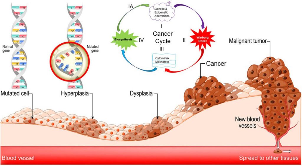
- Immune evasion Tumor cells suppress immune responses, enabling unchecked growth — the reason immunotherapies like Keytruda and Opdivo work.
For deeper reading, you can link to this authoritative review:
- Molecular pathways and therapeutic targets in lung cancer https://pmc.ncbi.nlm.nih.gov/articles/PMC4039220/
2. Medicines Used Today (General Mechanisms)
To organize modern lung cancer treatments, we use authoritative oncology sources rather than general drug catalogs. These include
- NCCN Guidelines (U.S.) — gold standard for oncology treatment
- ESMO Guidelines (Europe) — widely used across international oncology centers
- FDA Approvals (U.S.) — official regulatory decisions for cancer drugs
- EMA Approvals (Europe) — European regulatory authority
This ensures our analysis focuses on widely used, globally relevant medications with strong brand identity, measurable public interest, and clear molecular mechanisms.
| Drug Category | General Mechanism | Molecular Target / Pathway | Representative Brands |
|---|---|---|---|
| Immunotherapy (PD‑1 / PD‑L1 inhibitors) | Boost immune system recognition of cancer cells | PD‑1 / PD‑L1 checkpoint blockade | Keytruda, Opdivo, Tecentriq, Imfinzi |
| EGFR inhibitors | Block abnormal EGFR signaling that drives tumor growth | EGFR mutations (exon 19 del, L858R, T790M) | Tagrisso, Tarceva, Iressa, Gilotrif |
| ALK inhibitors | Inhibit ALK fusion proteins that cause uncontrolled proliferation | ALK rearrangements (EML4‑ALK) | Alecensa, Xalkori, Lorbrena, Alunbrig |
| KRAS G12C inhibitors | Target mutant KRAS signaling | KRAS G12C mutation | Lumakras, Krazati |
| ROS1 inhibitors | Block ROS1 fusion proteins | ROS1 rearrangements | Rozlytrek, Xalkori |
| RET inhibitors | Inhibit RET fusion‑driven signaling | RET rearrangements | Retevmo, Gavreto |
| MET inhibitors | Block MET amplification or exon 14 skipping | MET alterations | Tabrecta, Tepmetko |
| Chemotherapy backbone | Damage DNA or disrupt cell division | Non‑specific cytotoxic mechanisms | Carboplatin, Cisplatin, Paclitaxel, Pemetrexed |
3. APIs (Active Pharmaceutical Ingredients)
Active Pharmaceutical Ingredients (APIs) are the chemical or biological molecules that make cancer treatments work. Unlike brand names, APIs reveal the true identity of each therapy — its molecular target, mechanism of action, and scientific role in lung cancer treatment. Understanding APIs helps readers see how modern oncology is organized: each drug belongs to a molecular category such as immunotherapy, EGFR inhibitors, ALK inhibitors, KRAS inhibitors, ROS1 inhibitors, MET inhibitors, RET inhibitors, or chemotherapy.
These categories reflect the major pathways and mutations that drive tumor growth. They also align with globally recognized guidelines from NCCN, ESMO, FDA, and EMA, ensuring that the treatments shown here represent the most current standards of care.
Inside each API section (+), you will find: • A brief explanation of what the API is • How the drug works (mechanism of action) • Generations or evolution (if applicable) • The list of the most important APIs in that category • A racing chart or Google Trend showing recent interest • Reddit Insights • Twitter (X) Insights • Chemistry molecule visuals for key drugs
Click the + to expand each category.
What ALK inhibitors are
Anaplastic Lymphoma Kinase (ALK) inhibitors are targeted cancer therapies designed for patients whose tumors carry an ALK fusion, a genetic alteration that drives uncontrolled cell growth. These fusions occur in a small percentage of lung cancers, often in younger non‑smokers, and create an abnormal protein that acts like a stuck accelerator pedal for tumor growth.
How ALK inhibitors work
ALK inhibitors bind to the ATP‑binding pocket of the ALK fusion protein, shutting down the signaling pathways that tell cancer cells to survive and multiply. By blocking this abnormal signal, the drugs slow or stop tumor growth and often shrink tumors dramatically. Modern ALK inhibitors are engineered to reach the brain effectively, protecting patients from brain metastases — a common problem in ALK‑positive lung cancer.
Generations of ALK inhibitors
ALK inhibitors are grouped into three generations. Crizotinib, the first‑generation drug, was the original breakthrough but has limited brain penetration and resistance develops quickly. Second‑generation inhibitors (alectinib, brigatinib, ceritinib) are more potent and offer strong protection in the brain. Third‑generation lorlatinib is designed to overcome nearly all known resistance mutations and provides the most durable long‑term control.
| API | Brand Name(s) | Mutation | Mechanism |
|---|---|---|---|
| Alectinib | Alecensa | ALK fusion | Blocks ALK signaling |
| Crizotinib | Xalkori | ALK / ROS1 | Inhibits ALK & ROS1 |
| Lorlatinib | Lorbrena | ALK | Third‑generation ALK inhibitor |
| Brigatinib | Alunbrig | ALK | Inhibits ALK variants |
Chromatographic Methods Used to Quantify Lorlatinib
The table below summarizes validated quantitative HPLC methods for lorlatinib. The main scope is HPLC-UV/PDA; LC–MS/MS bioanalytical assays are excluded. The chiral method is listed separately because it evaluates enantiomeric purity rather than conventional tablet assay.
| Reference | Column | λ (nm) | Flow (mL/min) | Mobile phase | tR (min) | Linearity | Accuracy | Precision |
|---|---|---|---|---|---|---|---|---|
| Chakravarthi & Himabindu, 2021, Int. J. Res. Publ. Rev., 2(9):958–963 [1] | Eclipse Plus C18, 250 × 4.6 mm, 3 µm | 310 | 1.0 | Phosphate buffer pH 5.8 : ACN : MeOH, 45:30:25 v/v/v, isocratic | 7.871 | 50–150 µg/mL; r² = 0.9994 | Mean recovery 100.5%; individual recoveries 98.6–101.5% | System %RSD 0.1%; intermediate precision 0.3%; method precision 0.5% |
| Illendula & Sharma, 2024, Zhongguo Ying Yong Sheng Li Xue Za Zhi, 40:e20240009 [2][3] | Hypersil C18, 150 × 4.6 mm, 5 µm; 38 °C | 310 | 1.0 | MeOH : water, 25:75 v/v | 3.513 | NR* | Recovery 98.96% | Repeatability and intermediate precision reported as within limits; numerical values NR* |
| Liao et al., 2023, “Direct Enantioseparation of Lorlatinib Enantiomers by Liquid Chromatography on a Chiralpak IB Column,” J. Chem. Soc. Pak., 45(6):53 [4][5] | Chiralpak IB, 250 × 4.6 mm, 5 µm; 30 °C | 260 | 1.0 | n-Hexane : 2-propanol : diethylamine, 85:15:0.1 v/v/v | <10 | 10–1000 µg/mL, n = 8 | 97.86–100.99% recovery | RSD <1.6%; enantiomeric resolution >2.2 |
Notes
- NR = not reported numerically in the accessible source record.
- The 2021 paper contains an internal inconsistency: different sections report mobile-phase ratios of 45:30:25 and 50:30:20, and buffer pH values of 5.8 and 4.2. The table uses the optimized condition stated in the method-development/results section—phosphate buffer pH 5.8 : ACN : MeOH, 45:30:25—but this should be checked against the original chromatogram before laboratory adoption.[1]
- The Chiralpak IB method is a chiral-enantiomeric-purity method, not a conventional tablet assay method.
Sources
[1] https://ijrpr.com/uploads/V2ISSUE9/IJRPR1359.pdf
[2] https://pubmed.ncbi.nlm.nih.gov/39160080
[3] https://doi.org/10.62958/j.cjap.2024.009
[4] https://doi.org/10.52568/001195/jcsp/45.01.2023
[5] https://jcsp.org.pk/PublishedVersion/36edda5b-ec25-4c64-af05-e686f4bd930dManuscript%20no%209%2C%20Final%20Galley%20Proof%20of%2012638%20%28Qin%20Wang%29.pdf
ALK Reddit Insights
| API + Brand | Positive Themes | Negative Themes | Common Patient Discussions |
|---|---|---|---|
| Lorlatinib (Lorbrena) | – Extremely potent, often described as “king/queen of TKIs” – Rapid tumor shrinkage, including brain mets – Effective after failure of other TKIs | – Cognitive effects (“brain fog”, memory issues) – Edema, joint pain, high cholesterol – Shortness of breath (sometimes severe) | – Dose reductions (200 → 75 → 50 mg) to manage side effects – Switching to Lorlatinib after Alectinib/Brigatinib progression – Concerns about neuro side effects vs. strong efficacy |
| Alectinib (Alecensa) | – Very good tolerability for many patients – Strong efficacy, including complete responses in brain mets – Stable long‑term disease control | – Fatigue, muscle aches, sun sensitivity – Weight gain – Elevated liver enzymes | – First‑line treatment discussions – Comparisons with Lorlatinib (“slightly less potent but easier to tolerate”) – Stories of rapid improvement (“30% shrinkage in 3 days”) |
| Crizotinib (Xalkori) | – Early positive responses in some patients – Historically important first‑generation ALK inhibitor | – Vision disturbances (“light trails”) – GI issues – Lower efficacy vs. newer TKIs | – Often used as a control arm in studies – Many patients switching to Alectinib or Lorlatinib – Discussions about resistance developing |
| Brigatinib (Alunbrig) | – Good tolerance after initial adjustment period – Effective for some patients after Crizotinib failure | – Early pulmonary symptoms – Some cases of progression after long use | – Switching from Brigatinib to Lorlatinib after progression – Experiences of young patients with aggressive disease – Monitoring lung symptoms closely in first weeks |
ALK Twitter(X) Insights
| Drug | Effectiveness | Side Effects | Management / Community Insights |
|---|---|---|---|
| Lorlatinib | – Long‑term control of ALK‑positive lung cancer (7‑year data) – Strong protection against new brain tumors – Considered the most durable ALK treatment | – Some patients report memory, mood, or concentration issues | – Doctors share guides on managing side effects – Many discussions around choosing lorlatinib first‑line |
| Alectinib | – Many patient stories of long‑term remission – Effective after surgery (ALINA trial) | – Generally mild side effects | – Advocacy groups highlight its availability and tolerability |
| Crizotinib | – Important first‑generation ALK drug – Often used as comparison in new studies | – Less effective in the brain compared to newer drugs | – Mostly historical discussions; used as baseline comparator |
| Brigatinib | – Strong second‑generation option – Mentioned in first‑line choices in Europe | – Some tolerability trade‑offs | – Educational threads from advocacy groups |
What immunotherapy APIs are
Immunotherapy APIs are biological molecules designed to help the immune system recognize and attack cancer cells. In lung cancer, the most important immunotherapy drugs target the PD‑1/PD‑L1 pathway — a “shield” tumors use to hide from immune cells. By blocking this shield, immunotherapy reactivates the body’s natural defenses, allowing T‑cells to detect and destroy cancer more effectively. These treatments have transformed outcomes for many patients with advanced or early‑stage disease.
How immunotherapy works
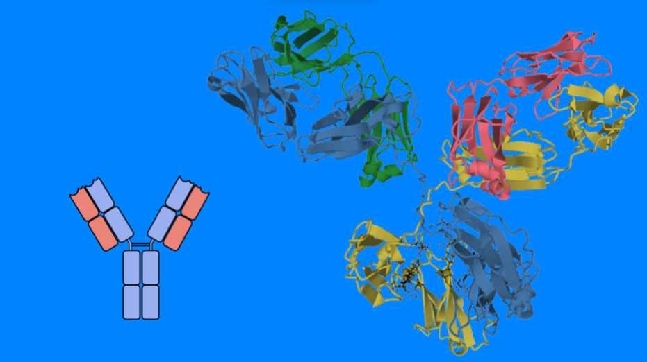
PD‑1 inhibitors (like pembrolizumab and nivolumab) and PD‑L1 inhibitors (like atezolizumab, durvalumab, and cemiplimab) bind to immune checkpoint proteins that normally act as brakes on the immune system. When cancer exploits these brakes, immune cells become inactive. Immunotherapy releases those brakes, restoring T‑cell activity and enabling long‑lasting immune responses. In some patients, this leads to durable control of the disease, even years after treatment stops.
Generations and evolution of immunotherapy
Immunotherapy does not have “generations” like ALK or EGFR inhibitors, but it has evolved through major clinical milestones. First came single‑agent PD‑1/PD‑L1 inhibitors, then combination strategies with chemotherapy, CTLA‑4 inhibitors, and targeted therapies. More recently, immunotherapy has moved into early‑stage lung cancer (neoadjuvant and adjuvant settings), and new approaches such as personalized mRNA cancer vaccines are emerging. These advances continue to expand the role of immunotherapy across multiple cancer types.
| API | Brand Name(s) | Target | Mechanism |
|---|---|---|---|
| Pembrolizumab | Keytruda | PD‑1 | Immune checkpoint blockade |
| Nivolumab | Opdivo | PD‑1 | Restores T‑cell activity |
| Atezolizumab | Tecentriq | PD‑L1 | Blocks PD‑L1 suppression |
| Durvalumab | Imfinzi | PD‑L1 | Enhances immune recognition |
| Cemiplimab | Libtayo | PD‑1 | Immune checkpoint inhibition |
Immunotherapy Twitter(X) Insights
| Drug / Topic | Effectiveness Signals | Side Effect / Concern Signals | Management / Community Insights |
|---|---|---|---|
| Pembrolizumab (Keytruda) | – Major 5‑year survival benefits in lung cancer (KEYNOTE‑671) – Strong results when combined with chemo in early‑stage NSCLC – Huge attention due to personalized mRNA vaccine + Keytruda success | – Some skepticism about cost and magnitude of benefit | – Many educational threads from oncologists – Discussions around biomarkers (PD‑L1) and who benefits most |
| mRNA personalized cancer vaccine + Keytruda | – First successful Phase 3 for an mRNA cancer vaccine – Reduced recurrence and distant metastases in melanoma – Considered a “landmark moment” by oncologists | – Concerns about cost and access – Questions about long‑term side effects | – Mechanism explained in simple threads (tumor sequencing → personalized vaccine) – Many posts about future trials in lung, kidney, bladder cancer |
| Nivolumab (Opdivo) | – Dual‑IO (Nivolumab + Ipilimumab) shows strong results in PD‑L1‑negative NSCLC – Some real‑world data suggests longer survival vs Keytruda + chemo in certain subgroups | – Higher toxicity when combined with Ipilimumab | – Discussions about choosing dual‑IO vs single‑agent IO + chemo |
| Atezolizumab (Tecentriq) | – Used in small‑cell lung cancer maintenance – Mentioned in comparisons with emerging agents | – Less frequent toxicity discussions | – Appears mainly in SCLC‑focused threads |
| Durvalumab (Imfinzi) | – Standard consolidation therapy for Stage III NSCLC – Strong long‑term control after chemoradiation | – Some posts mention fatigue | – Often referenced in treatment sequencing discussions |
| Cemiplimab (Libtayo) | – Effective in certain lung cancer and skin cancer settings | – Fewer mentions overall | – Appears in broader immunotherapy comparisons |
Immunotherapy Reddit Insights
| API + Brand | Positive Themes | Negative Themes | Common Patient Discussions |
|---|---|---|---|
| Pembrolizumab (Keytruda) | – Significant tumor shrinkage – Better tolerated than chemo – Long‑term remission cases | – Fatigue, joint pain, flu‑like symptoms – Colitis, diarrhea – Brain fog, sleep issues | – Managing colitis with steroids – Combination therapy with chemo – Individual variability |
| Nivolumab (Opdivo) | – Strong effectiveness in melanoma & lung cancer – Good outcomes with Yervoy combo – Long‑term stability | – Autoimmune reactions (colitis, thyroid) – Joint pain, arthritis – Fatigue | – Steroid management – Monitoring new symptoms – Combo therapy decisions |
| Atezolizumab (Tecentriq) | – Durable benefit in SCLC & NSCLC – Mild side effects for many – Long‑term disease control | – Vitiligo, nosebleeds, fatigue – Rare severe toxicity | – Distinguishing side effects vs progression – NCCN guideline checks – Second opinions |
| Durvalumab (Imfinzi) | – Tumor shrinkage after chemoradiation – Long‑term remission cases – Stabilization of disease | – Severe fatigue, appetite loss – Pneumonitis – Immune system overdrive | – Mixed responses – Emergency symptoms (O2 drop) – Individual variability |
What EGFR inhibitors are
EGFR inhibitors are targeted therapies designed for patients whose tumors carry specific mutations in the Epidermal Growth Factor Receptor (EGFR) gene. These mutations cause the receptor to remain permanently “on,” driving continuous cell growth and division. EGFR‑mutant lung cancer is one of the most common molecular subtypes, especially in non‑smokers and certain populations, making EGFR inhibitors a cornerstone of modern lung cancer treatment.
How EGFR inhibitors work
EGFR inhibitors bind to the ATP‑binding pocket of the mutated EGFR protein, blocking the signals that tell cancer cells to grow and survive. By shutting down this abnormal pathway, the drugs can shrink tumors rapidly and control disease for long periods. Third‑generation EGFR inhibitors are engineered to penetrate the brain effectively and to overcome resistance mutations such as T790M, which often appear after earlier treatments. Some EGFR inhibitors also work against rare mutations like exon 20 insertions, expanding treatment options for patients with less common tumor profiles.
Generations of EGFR inhibitors
EGFR inhibitors are grouped into three generations. First‑generation drugs (erlotinib, gefitinib) were the original breakthroughs but often led to resistance and had limited activity against certain mutations. Second‑generation inhibitors (afatinib, dacomitinib) are more potent and can target a broader range of EGFR alterations, though sometimes with increased side effects. Third‑generation osimertinib is now the preferred standard due to its strong effectiveness, excellent brain penetration, and ability to overcome resistance mutations. Newer agents for exon 20 insertions (like amivantamab and sunvozertinib) represent the next evolution of EGFR‑targeted therapy.
| API | Brand Name(s) | Mutation | Mechanism |
|---|---|---|---|
| Osimertinib | Tagrisso | EGFR T790M / L858R | Blocks mutant EGFR |
| Erlotinib | Tarceva | EGFR | Inhibits EGFR signaling |
| Gefitinib | Iressa | EGFR | Reduces EGFR proliferation |
| Afatinib | Gilotrif | EGFR / HER2 | Irreversible ErbB inhibition |
Molecule Profile
| Property | Value |
|---|---|
| Molecule | Osimertinib |
| Brand Name | Tagrisso |
| Company | AstraZeneca |
| Formula | C₂₈H₃₃N₇O₂ |
| Molecular Weight | 499.6 g/mol |
| Drug Class | EGFR Tyrosine Kinase Inhibitor |
| Chemical Class | Aminopyrimidine |
| Generation | Third Generation |
Chromatographic Methods Used to Quantify Osimertinib
The table below summarizes validated quantitative HPLC methods for osimertinib that were identifiable in the retrieved literature.[1][3] The main scope is HPLC with UV/PDA detection for bulk drug or pharmaceutical formulations; LC–MS/MS bioanalytical assays are excluded. Two additional HPLC methods are included with an explicit evidence limitation because their chromatographic parameters were accessible through a later comparison table, while the primary full texts were not retrieved in this pass.[3]
| Reference | Column | λ (nm) | Flow (mL/min) | Mobile phase | tR (min) | Linearity | Accuracy | Precision |
|---|---|---|---|---|---|---|---|---|
| Alhabardi, Aljohar, Alganamy & Almurshedi, 2025, Acta Chromatographica, 38(2):376–386 [3][4] | Nova-Pak C18, 250 × 3.9 mm, 4 µm; 29 °C | 276 | 1.0 | ACN : 0.1 M ammonium acetate, 1:1 (v/v), isocratic | NR in the accessible article text | 2.40–20.29 µg/mL (R² = 0.9988) | 90.034–105.227%; mean recoveries 102.9%, 104.83%, and 90.164% at 4, 8, and 16 µg/mL | Intra-day %RSD 0.192–0.472%; inter-day %RSD 0.28–0.47% |
| Akter, Rajbangshi, Faruk, Shill & Rouf, 2024, Bangladesh Pharmaceutical Journal, 27(1):85–91 [1][2] | Prontosil C18, 150 × 4.6 mm i.d., 3.5 µm; 30 °C | 283 | 0.7 | Triethylamine buffer (pH 2.5) : ACN, 65:35 (v/v), isocratic | 3.640 ± 0.28 | 64–96 µg/mL (R² > 0.999) | Tablet recovery 99.32–100.37% (mean ± %RSD at levels: 100.12 ± 0.11%, 99.32 ± 0.42%, 100.37 ± 0.42%) | Intra-day tablet recovery 100.03 ± 0.40%; inter-day mean 99.78 ± 0.61% |
| Shelar Shweta, Mateen, Iqbal et al., 2023, Journal of Chemical Health Risks, 13(2) [3] | NR in the accessible source record | 270 | 1.0 | Perchloric acid : MeOH, 80:20 (v/v) | 3.28 | NR in the accessible source record | NR in the accessible source record | NR in the accessible source record |
| Bhutnar, Sapale, Vaidya & Desai, 2022, International Journal of Pharmaceutical Sciences and Drug Research, pp. 739–748 [3][5] | NR in the accessible source record | NR | 1.0 | Gradient: 0.1% formic-acid buffer (pH 6.50) : ACN; diluent = mobile phase A : ACN, 1:1 (v/v) | OSM: 41.76; impurities: 25.99, 29.87, 32.90, 33.67, 39.15, 35.73 | NR in the accessible source record | NR in the accessible source record | NR in the accessible source record |
| Chakradhar, Mondal & Vanapalli, 2019, Journal of Drug Delivery & Therapeutics, 9(2-s):391–395 [3][6] | NR in the accessible source record | 265 | 1.0 | Gradient: 0.1 M phosphate buffer (pH 3.0) : ACN, 70:30 → 50:50 (v/v) | 5.20 ± 0.15 | NR in the accessible primary source record | NR in the accessible primary source record | NR in the accessible primary source record |
Notes
- NR = not reported numerically in the accessible source record; it should not be interpreted as absent from the full paper.
- The 2024 Akter et al. method is the most completely documented in the retrieved primary text: it reports a PDA detector, 15 µL injection, 10-minute run time, C18 dimensions, wavelength, retention time, linearity, recovery, precision, LOD (0.4 µg/mL), and LOQ (1.2 µg/mL).[1]
- The 2025 Alhabardi et al. paper reports the final chromatographic conditions and validation results directly, but its accessible text contains internal inconsistencies: the materials section states 1 mL/min, while a later results passage states 0.1 mL/min; the abstract/method conditions state 276 nm, while one figure caption states 213 nm. The table uses the conditions stated in the abstract/materials method description (1 mL/min, 276 nm), but the original PDF should be checked before laboratory adoption.[3]
- The 2025 paper’s recovery table reports individual recoveries of 103.006–103.021–102.671% at 4 µg/mL, 105.227–104.988–104.277% at 8 µg/mL, and 90.034–90.024–90.434% at 16 µg/mL; the table above reports the paper’s summarized ranges and means.[3]
- The 2019, 2022, and 2023 rows use chromatographic parameters reproduced in Table 1 of the 2025 paper, which identifies the primary references as Chakradhar et al., Bhutnar et al., and Shelar Shweta et al., respectively.[3][5][6] Their numeric validation fields are marked NR because the primary full texts were not accessible in this retrieval pass.
- LC–MS/MS methods for osimertinib in plasma, cerebrospinal fluid, and other biological matrices were not included because the reference format is centered on HPLC-UV/PDA quantitative methods for bulk drug and formulations.[1][3]
Sources
[1] https://banglajol.info/index.php/BPJ/article/download/71158/47788
[2] https://doi.org/10.3329/bpj.v27i1.71158
[3] https://www.akjournals.com/view/journals/1326/38/2/article-p376.xml
[4] https://doi.org/10.1556/1326.2025.01379
[5] https://doi.org/10.25004/IJPSDR.2022.140611
[6] https://doi.org/10.22270/jddt.v9i2-s.2733
Protein Profile
| Property | Value |
|---|---|
| Protein | EGFR |
| PDB ID | 7U9A |
| Organism | Homo sapiens |
| Classification | Transferase / Transferase Inhibitor |
| Protein Mass | 38.16 kDa |
| Atom Count | 2417 |
| Modeled Residues | 295 |
| Unique Protein Chains | 1 |
| Resolution | 2.60 Å |
| Method | X-Ray Diffraction |
Scientific & Community Interest
Google Trends
EGFR X (Twitter)
| Drug / Topic | Effectiveness Signals | Side Effect / Concern Signals | Management / Community Insights |
|---|---|---|---|
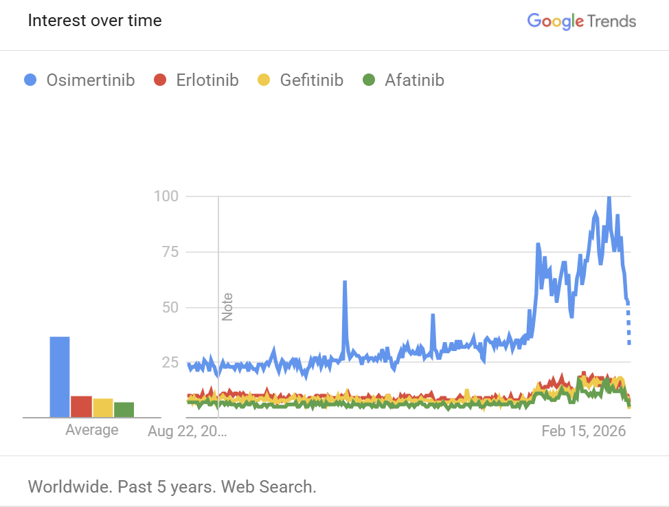
| Osimertinib (Tagrisso) | – Dominates EGFR discussions on Twitter – Strong long‑term control in early‑stage lung cancer (ADAURA updates) – Promising results in resistance settings when combined with other drugs (HARMONi, ORCHARD, SAFFRON) – Many patient stories of major tumor shrinkage | – Some posts mention resistance mutations (C797X, MET amplification, RAS changes) | – Oncologists share educational threads on EGFR mutation types – Discussions on sequencing after resistance – High engagement around new trial results |
| Erlotinib (Tarceva) | – Mostly referenced historically – Appears in older sequencing strategies | – More side effects compared to newer TKIs (mentioned indirectly) | – Used as comparison point in expert discussions |
| Gefitinib (Iressa) | – Mentioned in niche trials (e.g., adjuvant gefitinib + chemo) – Appears in combination discussions for resistance (osimertinib + gefitinib) | – Less effective than osimertinib in modern practice | – Mostly referenced in resistance or historical contexts |
| Afatinib (Gilotrif) | – Occasionally mentioned for rare EGFR mutations | – Known for stronger side effects (skin/GI), mentioned indirectly | – Appears in discussions about older TKIs and niche mutation coverage |
EGFR Reddit
| API + Brand | Positive Themes | Negative Themes | Common Patient Discussions |
|---|---|---|---|
| Osimertinib (Tagrisso) | – Rapid initial improvement (first 10 days) – Long‑term disease control for many – Effective for brain mets and resistant mutations | – Skin issues (rash, rosacea, cracked skin) – Fatigue – GI issues (diarrhea, nausea, appetite loss) | – Resistance development (“progression after 2 years”) – Adding chemo after resistance (FLAURA2) – Managing skin toxicity (minocycline, moisturizers) – Hydration strategies |
| Erlotinib (Tarceva) | – Effective for certain EGFR mutations – Some patients achieve stable disease – Can work well when mutation profile matches | – Severe skin rash (acneiform) – GI issues (diarrhea, nausea) – Fatigue, appetite loss – Mouth sores (mucositis) | – Genetic factors affecting response – Dose adjustments for rash/diarrhea – Balancing quality of life vs effectiveness – Symptomatic management (creams, anti‑diarrheals) |
| Afatinib (Gilotrif) | – Effective for specific EGFR mutations (G719D, exon 19 variants) – Side effects often stabilize over time – Dose reduction maintains benefit | – Diarrhea (very common) – Skin rash, pimples, dry skin – Throat pain, mucositis, dry cough | – Hydration + electrolytes for diarrhea – Skin care routines (moisturizers, sunscreen, steroids) – Gargling + humidifier for throat pain – Dose reduction strategies |
| Gefitinib (Iressa) | – Tumor shrinkage in some NSCLC cases – Stable disease or remission when combined with immunotherapy | – Extreme fatigue (“slept 23 hours/day”) – Severe GI issues (vomiting, diarrhea) – Rash, itching, mouth sores – Appetite loss, weight loss | – Using Zofran for nausea – Steroids for inflammation – Dietary adjustments (cold foods, low acid) – Communicating side effects to oncologists |
What KRAS G12C inhibitors are
KRAS G12C inhibitors are targeted therapies designed for tumors carrying the KRAS G12C mutation — one of the most common and historically “undruggable” drivers of lung cancer and colorectal cancer. KRAS mutations act like a stuck accelerator pedal, keeping cancer cells in a constant growth mode. The G12C variant creates a unique cysteine residue that allows modern drugs to bind and lock KRAS in its inactive state, making this mutation finally treatable after decades of failed attempts.
How KRAS G12C inhibitors work
KRAS G12C inhibitors bind to the mutant KRAS protein in its inactive GDP‑bound form, trapping it so it cannot switch back into the active signaling state. By shutting down this pathway, the drugs reduce downstream MAPK signaling — the chain reaction that drives tumor growth and survival. In lung cancer, this often leads to meaningful tumor shrinkage and improved disease control. In colorectal cancer, KRAS G12C inhibitors are typically combined with EGFR inhibitors because CRC tumors rapidly reactivate growth pathways unless EGFR is blocked at the same time.
Generations and evolution of KRAS G12C inhibitors
KRAS G12C inhibitors do not have formal “generations” yet, but the field is evolving rapidly. The first approved agents — sotorasib and adagrasib — opened the door to targeting KRAS directly. Newer, next‑generation inhibitors such as divarasib are showing stronger potency and improved activity in early trials, especially in pretreated lung cancer. Combination strategies (KRAS + EGFR inhibitors, KRAS + SHP2 inhibitors, KRAS + immunotherapy) represent the next wave of development, aiming to overcome resistance and improve outcomes across multiple cancer types.
| API | Brand Name(s) | Mutation | Mechanism |
|---|---|---|---|
| Sotorasib | Lumakras | KRAS G12C | Irreversible KRAS inhibition |
| Adagrasib | Krazati | KRAS G12C | Blocks mutant KRAS signaling |
Chromatographic Methods Used to Quantify Sotorasib
The table summarizes quantitative HPLC methods for sotorasib identified in the retrieved literature.[1][2] The main scope is HPLC with UV/PDA detection. The methods differ in matrix and intended use: the 2021 method is for pharmaceutical/API analysis, whereas the 2023 method is for plasma therapeutic-drug monitoring. A chiral atropisomer-separation application note is included separately.[3]
| Reference | Column | λ (nm) | Flow (mL/min) | Mobile phase | tR (min) | Linearity | Accuracy | Precision |
|---|---|---|---|---|---|---|---|---|
| Yarlagadda, Mannam & Jampani, 2021, International Journal of Applied Pharmaceutics, 13(5):154–159 [1] | Symmetry C18, 150 × 4.6 mm, 3.5 µm; PDA detector | 221 | 1.0 | Acetonitrile : 0.1% orthophosphoric acid, 70:30 (v/v), isocratic | 2.271 | 10–150 µg/mL (r² 0.99982) | Article text reports recovery 99.38–99.71%; accuracy/assay results were within the reported acceptance range | Intraday %RSD 0.66%; interday %RSD 0.765%; six-replicate system %RSD about 0.59% |
| Hikita et al., 2023, Drug Discoveries & Therapeutics, 17(4):289–293 [2] | Capcell Pak C18 MG II, 250 × 4.6 mm, 5 µm, with 10 × 4.0 mm guard column | 230 | 1.0 | 0.5% KH₂PO₄, pH 4.5 : acetonitrile, 65:35 (v/v), isocratic | 14.5 (sotorasib); 10.8 (bosutinib IS) | 0.10–20.0 µg/mL (r² 0.9999) | Intra-day accuracy −3.88 to +5.18%; inter-day accuracy −3.14 to −0.39%; recovery 95.83–106.92% | Intra-day CV 0.79–9.75%; inter-day CV 3.01–6.13% |
Separate chiral / atropisomer method
| Reference | Column | λ (nm) | Flow (mL/min) | Mobile phase | tR (min) | Linearity | Accuracy | Precision |
|---|---|---|---|---|---|---|---|---|
| Daicel Chiral Technologies, “The Chiral Analytical and Preparative Resolution of Sotorasib Atropisomers,” application note [3] | CHIRALPAK IK, 250 × 4.6 mm, 5 µm; an IE method was also optimized | 305 | 1.0 | Acetonitrile; sample prepared in ACN:MeOH, 2:1 (v/v) | NR | NR | NR | NR |
Notes
- NR = not reported numerically in the accessible source.
- The 2021 method is an achiral, stability-indicating RP-HPLC/PDA assay for sotorasib API/pharmaceutical dosage-form analysis.[1]
- The 2023 Hikita method is a bioanalytical HPLC-UV plasma assay for therapeutic drug monitoring, not a dosage-form assay. Bosutinib was used as the internal standard.[2]
- The 2021 paper contains an internal inconsistency in its recovery presentation: the prose reports 99.38–99.71%, whereas the accessible OCR of Table 3 shows values around 99.7–101.3%. This should be checked against the original published table before use.[1]
- The Daicel note reports chromatographic conditions for analytical/preparative atropisomer separation but does not provide a comparable validation range, recovery, or precision dataset.[3]
- LC-MS/MS methods were excluded from the main table to match the scope of the previous Lorlatinib table.[4]
Sources
[1] https://journals.innovareacademics.in/index.php/ijap/article/download/42659/25336
[2] https://www.jstage.jst.go.jp/article/ddt/advpub/0/advpub_2023.01043/_pdf
[3] https://chiraltech.com/wp-content/uploads/2023/04/Daicel-Chiral-Technologies-Application-Note-Sotorasib-Atropisomers.pdf
[4] https://doi.org/10.1016/j.rechem.2024.101799
Google Trends
KRAS G12C Twitter (X) Insights
| Drug / Topic | Effectiveness Signals | Side Effect / Concern Signals | Management / Community Insights |
|---|---|---|---|
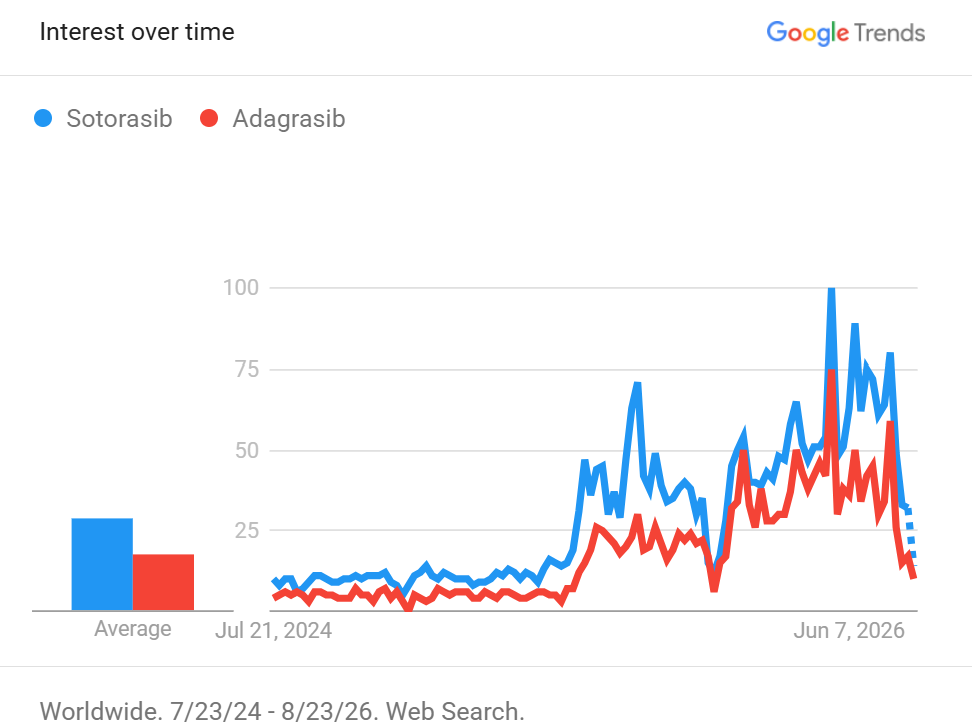
| Sotorasib (Lumakras) | – Strong results in colorectal cancer when combined with other drugs (e.g., panitumumab + FOLFIRI) – Good real‑world outcomes in advanced lung cancer | – High rates of severe side effects in CRC triplet combinations (≈50% grade ≥3) – Past concerns about liver toxicity when combined with immunotherapy | – Many posts about ongoing Phase III validation – Frequently discussed in CRC combination strategies |
| Adagrasib (Krazati) | – Promising CRC results when combined with cetuximab (some complete responses) – Quality‑of‑life maintained in KRYSTAL‑12 lung cancer study | – Resistance mechanisms discussed (e.g., EML4‑ALK fusion emerging after treatment) | – Oncologists share resistance explanations and sequencing strategies |
| Next‑generation KRAS inhibitors | – New agents (e.g., divarasib) show potential superiority over sotorasib/adagrasib in pretreated lung cancer | – Toxicity profiles still being evaluated | – Strong interest from researchers and AI/drug‑discovery communities |
| Broader KRAS G12C themes | – Clear differences between lung cancer vs colorectal cancer effectiveness – Combination therapy is the dominant strategy in CRC | – GI, liver, and skin toxicities frequently mentioned in combinations | – Heavy discussion around resistance mutations and how to overcome them |
KRAS G12C Reddit Insights
| API + Brand | Positive Themes | Negative Themes | Common Patient Discussions |
|---|---|---|---|
| Adagrasib (Krazati) | – Long‑term disease control (2+ years) – Shrinkage of tumors below diaphragm – Crosses blood‑brain barrier (prevents new mets) | – Persistent diarrhea (daily) – Nausea/vomiting early in treatment – Fatigue and bone pain | – Gradual dose increase to reduce vomiting – Using KRAS Kickers support group – Adjusting to chronic GI side effects – Monitoring brain mets risk |
| Sotorasib (Lumakras) | – Complete remission in some Stage 3 cases – Crosses blood‑brain barrier (prevents new mets) – Some patients tolerate it well | – Severe lung inflammation (rare but catastrophic) – Extreme diarrhea for weeks – Elevated liver enzymes – Extreme fatigue – Cases of rapid deterioration | – Financial assistance programs for cost – Distinguishing side effects vs progression – Variability (“tolerated well vs fatal toxicity”) – Monitoring liver enzymes and lung inflammation |
What ROS1 inhibitors are
ROS1 inhibitors are targeted therapies designed for patients whose tumors carry a ROS1 fusion, a rare but important driver of lung cancer. These fusions create an abnormal ROS1 protein that continuously sends growth signals to cancer cells, acting like a stuck accelerator. Although ROS1‑positive lung cancer is uncommon, it often responds extremely well to targeted therapy, making ROS1 inhibitors a crucial option for patients with this molecular subtype.
How ROS1 inhibitors work
ROS1 inhibitors bind to the ATP‑binding pocket of the ROS1 fusion protein, blocking the abnormal signaling pathways that promote tumor growth and survival. By shutting down this pathway, these drugs can shrink tumors rapidly and provide meaningful disease control. Some ROS1 inhibitors also target related proteins (such as ALK or TRK), and modern agents are engineered to penetrate the brain effectively — an essential feature because ROS1‑positive lung cancer can spread to the central nervous system.
Generations and evolution of ROS1 inhibitors
ROS1 inhibitors do not have formal “generations” like ALK or EGFR therapies, but the field has evolved through clear phases. Crizotinib and entrectinib were the first widely used ROS1 inhibitors, offering strong initial responses but limited durability against resistance mutations. Newer, next‑generation agents — including repotrectinib, zidesamtinib, and taletrectinib — are designed to overcome resistance and provide better CNS protection. These emerging therapies represent the future of ROS1‑targeted treatment, offering more potent and longer‑lasting control.
| API (Brand) | Mutation | Mechanism | Category | Functional Groups | Protein Target |
|---|---|---|---|---|---|
| Entrectinib (Rozlytrek) | ROS1 fusion | Blocks ROS1 signaling; also inhibits TRK (NTRK1/2/3); strong CNS penetration | ROS1 inhibitor | Indazole, piperazine | ROS1, TRK |
| Crizotinib (Xalkori) | ROS1 fusion | Blocks ROS1 signaling; also inhibits ALK and MET; limited CNS penetration | ROS1 inhibitor | Piperidine, aminopyridine | ROS1, ALK, MET |
| Repotrectinib (Augtyro) | ROS1 fusion | Highly potent ROS1 inhibition; designed to overcome resistance mutations; also inhibits TRK | ROS1 inhibitor | Cyclopropane, fluorinated aromatic rings, piperazine | ROS1, TRK |
ROS1 Twitter (X) Insights
| Drug / Topic | Effectiveness Signals | Side Effect / Concern Signals | Management / Community Insights |
|---|---|---|---|
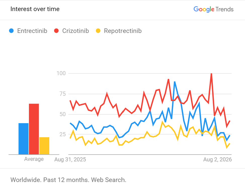
| Crizotinib (Xalkori) | – Often used as the historical baseline for ROS1 and ALK – Strong early activity but limited long‑term durability compared to newer drugs | – Lower protection for the brain compared to newer TKIs | – Frequently referenced in comparisons with next‑generation ALK/ROS1 drugs – Appears in discussions about resistance and sequencing |
| Entrectinib (Rozlytrek) | – Effective for ROS1+ lung cancer and NTRK‑positive tumors – Good brain penetration highlighted in scientific posts | – Some posts mention reimbursement/access issues in certain regions | – Discussions about newer ROS1 drugs overtaking entrectinib in some markets – Mentioned in educational threads for NTRK fusion cancers |
| Next‑generation ROS1 TKIs | – Strong interest in repotrectinib, zidesamtinib, taletrectinib – Newer agents expected to outperform crizotinib/entrectinib | – Still emerging toxicity profiles | – Many posts anticipate these drugs becoming the new standard – Access and reimbursement discussions appear frequently |
| Broader ROS1 themes | – ROS1 discussions often overlap with ALK due to shared drug history – CNS activity and resistance are major topics | – Resistance mechanisms (e.g., solvent‑front mutations) mentioned indirectly | – Lower tweet volume compared to EGFR or Immunotherapy – Posts cluster around trial updates and drug approvals |
ROS1 Reddit Insights
| API (Brand) | Mutation | Mechanism | Category | Functional Groups | Protein Target |
|---|---|---|---|---|---|
| Entrectinib (Rozlytrek) | ROS1 fusion | Blocks ROS1 signaling; also inhibits TRK (NTRK1/2/3); strong CNS penetration | ROS1 inhibitor | Indazole, piperazine | ROS1, TRK |
| Crizotinib (Xalkori) | ROS1 fusion | Blocks ROS1 signaling; also inhibits ALK and MET; limited CNS penetration | ROS1 inhibitor | Piperidine, aminopyridine | ROS1, ALK, MET |
What MET inhibitors are
MET inhibitors are targeted therapies designed for tumors driven by alterations in the MET gene, most commonly MET exon 14 skipping mutations or MET amplification. These abnormalities cause the MET receptor to remain constantly active, sending continuous growth and survival signals to cancer cells. MET‑altered lung cancer represents a distinct molecular subtype, and MET inhibitors provide an important treatment option for patients whose tumors rely on this pathway.
How MET inhibitors work
MET inhibitors bind to the MET receptor’s kinase domain, blocking the abnormal signaling that promotes tumor growth, survival, and metastasis. By shutting down this pathway, MET inhibitors can shrink tumors and slow disease progression. They are particularly effective in MET exon 14 skipping cancers, where the mutation prevents normal degradation of the MET protein, leading to excessive signaling. MET inhibitors are also used in combination with EGFR inhibitors when MET amplification emerges as a resistance mechanism after EGFR‑targeted therapy.
Generations and evolution of MET inhibitors
MET inhibitors do not have formal “generations,” but the field has evolved through clear phases. Early multi‑target drugs like crizotinib showed activity against MET, but modern selective MET inhibitors — such as capmatinib and tepotinib — provide stronger, more precise inhibition with better tolerability. Newer agents and combination strategies (MET + EGFR inhibitors, MET + antibody therapies) are being developed to overcome resistance and improve outcomes, especially in patients whose tumors develop MET amplification after EGFR therapy.
| API | Brand Name(s) | Mutation | Mechanism |
|---|---|---|---|
| Capmatinib | Tabrecta | MET exon 14 skipping | Blocks MET signaling |
| Tepotinib | Tepmetko | MET exon 14 skipping | Inhibits MET pathway |
MET Twitter (X) Insights
| Drug / Topic | Effectiveness Signals | Side Effect / Concern Signals | Management / Community Insights |
|---|---|---|---|
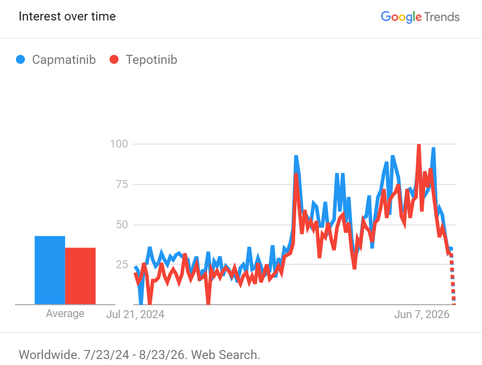
| Capmatinib (Tabrecta) | – Strong activity in MET exon 14 skipping lung cancer – Real‑world posts show meaningful tumor shrinkage | – Edema frequently mentioned (hands, legs, face) – Fatigue and GI issues appear in patient discussions | – Oncologists discuss dose adjustments for edema – Posts highlight monitoring kidney function and hydration |
| Tepotinib (Tepmetko) | – Similar effectiveness to Capmatinib in MET exon 14 skipping NSCLC – Some posts mention stable disease in real‑world use | – Edema also common; tolerability varies by patient | – Discussions around switching between MET TKIs if side effects are difficult |
| Broader MET themes | – MET amplification after EGFR therapy is a major topic – Many posts discuss combining MET inhibitors with EGFR TKIs (e.g., osimertinib + savolitinib) | – Combination therapy increases toxicity (fatigue, GI, edema) | – Oncologists share sequencing strategies for MET amplification after resistance to EGFR drugs |
MET Reddit Insights
| API + Brand | Positive Themes | Negative Themes | Common Patient Discussions |
|---|---|---|---|
| Capmatinib (Tabrecta) | – Significant tumor shrinkage (50% reduction reported) – Long‑term disease control – Improved quality of life | – Edema (hands, feet, legs) – Fatigue – Urinary issues from fluid retention | – Compression socks/sleeves for edema – Dose adjustments – Managing fatigue – Monitoring progression with scans |
| Tepotinib (Tepmetko) | No meaningful Reddit discussions found | No meaningful Reddit discussions found | No meaningful Reddit discussions found |
What RET inhibitors are
RET inhibitors are targeted therapies designed for tumors driven by RET fusions or RET mutations. These genetic alterations activate the RET receptor abnormally, sending continuous growth and survival signals to cancer cells. RET fusions are found in a small but important subset of lung cancers, while RET mutations are common in medullary thyroid cancer. Because RET‑altered tumors depend heavily on this pathway, selective RET inhibitors can produce strong and durable responses.
How RET inhibitors work
RET inhibitors bind to the kinase domain of the RET protein, blocking the abnormal signaling that drives tumor growth. By shutting down this pathway, these drugs can shrink tumors rapidly and provide long‑lasting disease control. Modern RET inhibitors are highly selective, meaning they target RET precisely while sparing many other kinases — a design that improves effectiveness and reduces side effects. They also penetrate the brain well, an essential feature for treating lung cancers that may spread to the central nervous system.
Generations and evolution of RET inhibitors
RET inhibitors do not have formal “generations,” but the field has evolved dramatically. Early multi‑target drugs like cabozantinib and vandetanib showed partial activity but were limited by off‑target toxicities. The development of highly selective RET inhibitors — selpercatinib and pralsetinib — transformed treatment outcomes, offering stronger responses with better tolerability. Selpercatinib has recently moved into early‑stage lung cancer (adjuvant therapy), marking a major milestone and positioning RET alongside EGFR and ALK as a validated early‑stage target.
| API | Brand Name(s) | Mutation | Mechanism |
|---|---|---|---|
| Selpercatinib | Retevmo | RET fusion | RET tyrosine kinase inhibition |
| Pralsetinib | Gavreto | RET fusion | Blocks RET signaling |
Chromatographic Methods Used to Quantify Selpercatinib
The pyrazolo[1,5-a]pyridine–pyridine core in the structure above is the UV chromophore that makes selpercatinib detectable by HPLC-UV/PDA, typically at 218–248 nm. The table lists only validated quantitative HPLC-UV/PDA methods published between 2021 and 2026 for which at least five of the nine columns are numeric. LC–MS/MS, NMR, and preparative methods are omitted. Retention time (tR) is that of the parent drug.
| Reference | Column | λ (nm) | Flow (mL/min) | Mobile phase | tR (min) | Linearity | Accuracy | Precision |
|---|---|---|---|---|---|---|---|---|
| Rajurkar et al., 2026, IJDDT 16(28s) | Inertsil C18, 250 × 4.6 mm, 5 µm | 238 | 1.0 | A: NaH₂PO₄ pH 4.0; B: MeOH; gradient 10→50% B | 12.66 | 10–50 µg/mL (r² 0.9993) | Recovery 99.2–99.9% | Intra-day %RSD 0.7%; intermediate 0.8% |
| Madhvilatha et al., 2025, Indian J. Pharm. Biol. Res. 13(2):1–7 | C18, 250 × 4.6 mm, 5 µm | 240 | 1.0 | 0.2% TFA pH 6.5–ACN 50:50 | 7.76 | 2–10 µg/mL (r² 0.9995) | Recovery 100.33–101.05% (mean 100.61%) | Repeatability %RSD 1.72% |
| Xiang et al., 2025, Front. Chem. 12:1534132 | C18, 250 × 4.6 mm, 5 µm, 35 °C | 235 | 1.0 | A: H₂O–ACN 9:1, pH 2.5, 2 mM KH₂PO₄, 0.4% TEA; B: ACN; gradient 5→45% B | 21.37 | 0.1–10 µg/mL (r 0.9994) | Impurity recovery 95.4–103.5% | Area %RSD 0.68–1.29%; repeatability 0.95–1.22% |
| Suzuki et al., 2024, Drug Discov. Ther. 18(6):387–390 | Capcell Pak C18 MG II, 250 × 4.6 mm | 240 | 1.0 | 0.5% KH₂PO₄ pH 4.5–ACN 70:30 | 8.3 | 0.5–8.0 µg/mL (r² 0.9996) | Recovery 94.43–97.30% | Intra- and inter-day CV ≤ 2.80% |
| Divya et al., 2024, FABAD J. Pharm. Sci. 49(3):449–464 | Zorbax C18, 250 × 4.6 mm, 5 µm, 35 °C | 226 | 0.9 | NH₄OAc–MeOH 70:30, pH 4.5, 0.1% FA (isocratic) | 8.41 | 15–120 µg/mL | — | — |
| Paka et al., 2024, J. Drug Deliv. Ther. 14(7) | Hypersil ODS C18, 250 × 4.6 mm, 5 µm | 248 | 1.0 | 0.2% TFA pH 6.5–ACN 70:30 | 3.01 | 2.5–15 µg/mL (r² 0.9993) | Mean recovery 100.59% | System %RSD 0.85%; method 0.47% |
| Nhavale et al., 2024, IJPQA 15(2) | Kromasil C18, 100 × 4.6 mm | 220 | 0.7 | MeOH–0.1% acetic acid 80:20, pH 3.0 | 3.20 | 10–50 µg/mL (r² 0.999) | Recovery 99.00–99.51% | Intra-day %RSD 0.00–0.10%; inter-day 0.11–0.16% |
| Katta & Thakre, 2024, Asian J. Chem. 36(2):341–352 | ZORBAX Eclipse, 250 × 4.6 mm, 5 µm, 35 °C | 241 | 0.8 | 0.5 M NaClO₄ pH 5.4–MeOH–ACN 50:20:30 (isocratic) | 7.34 | 30–240 µg/mL (r² 0.9991) | Recovery 99.44–99.51% | Intra-day %RSD 0.31%; inter-day 0.16–0.31% |
| Asok Kumar et al., 2024, IJSDR 9(1) | Kromosil C18, 250 × 4.6 mm, 5 µm, 30 °C | 218 | 1.0 | Na₂HPO₄–MeOH 55:45 | 2.33 | 150–6000 ng/mL (r² 0.999) | Mean recovery 99.52% | Intra-day %CV 0.25–11.45%; inter-day 0.35–0.85% |
| Singamsetty & Sundararajan, 2021, Int. J. Res. Pharm. Sci. 12(1):931–939 | Zorbax C18, 150 × 4.6 mm, 5 µm | — | 1.0 | 0.1% H₃PO₄–ACN 60:40 | 2.65 | 5–30 µg/mL (r 0.999) | Recovery 98.61–100.87% | %RSD 0.6 |
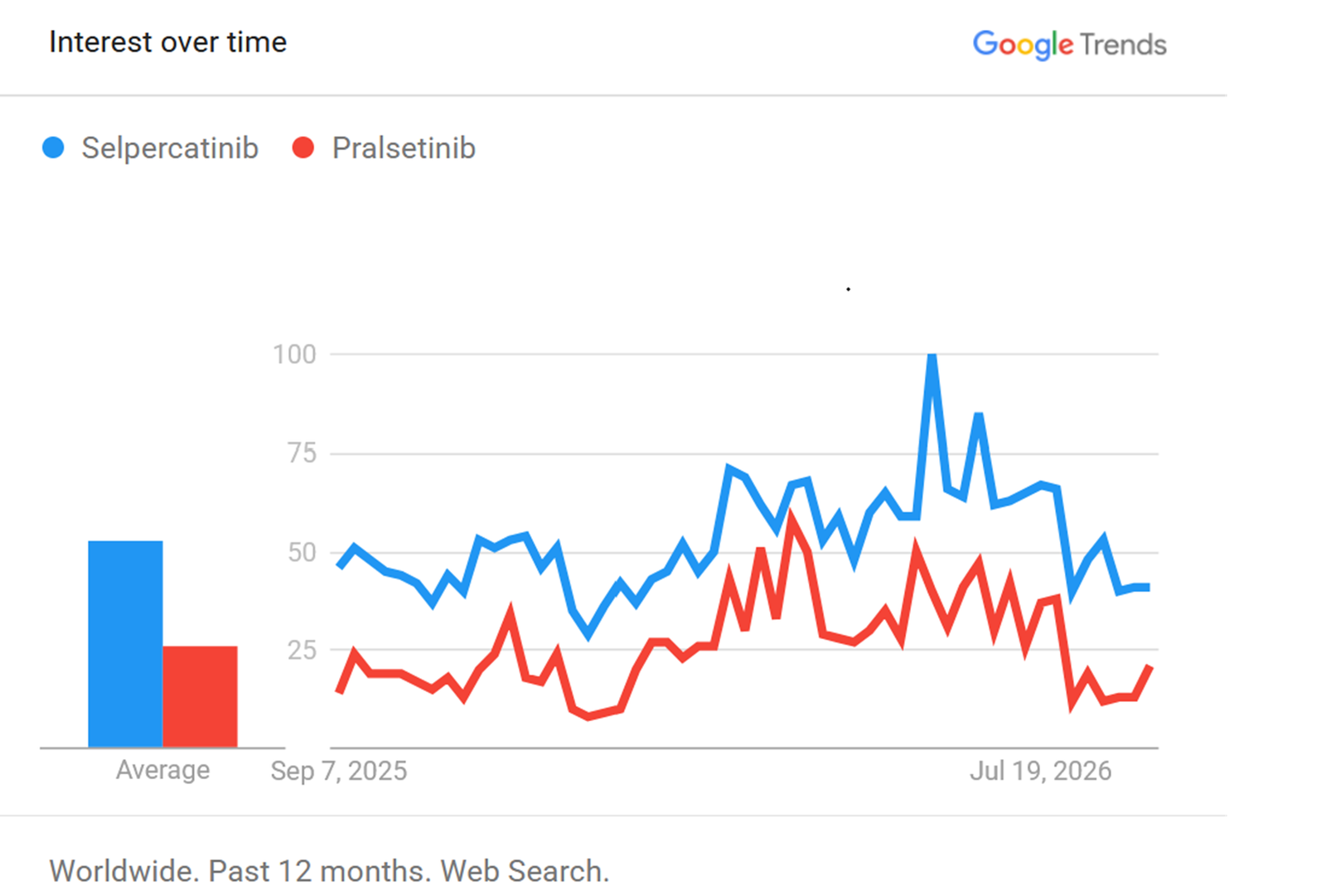
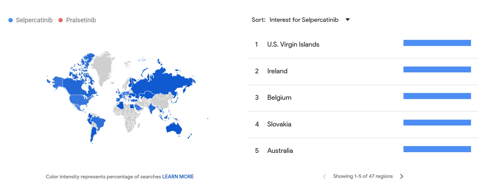
Google Trends
RET Twitter (X) Inhibitor APIs
| Drug / Topic | Effectiveness Signals | Side Effect / Concern Signals | Management / Community Insights |
|---|---|---|---|
| Selpercatinib (Retevmo) | – Very strong results in early‑stage RET‑positive lung cancer (LIBRETTO‑432) – 2‑year event‑free survival ~92% vs 61% with placebo – Durable responses in multiple RET‑altered cancers | – Elevated liver enzymes mentioned as the main Grade ≥3 toxicity | – Oncologists emphasize biomarker testing for RET fusions – Many posts highlight selpercatinib’s growing role in adjuvant therapy – Frequently listed among top ASCO 2026 lung cancer abstracts |
| Pralsetinib (Gavreto) | – High response rates in RET‑positive lung cancer (ARROW study) – Effective in both treatment‑naïve and pretreated patients | – Similar toxicity profile to selpercatinib (liver enzymes, mild GI) | – Often discussed as an alternative when selpercatinib is unavailable – Appears in guideline summaries and rare‑driver overviews |
| Broader RET themes | – RET therapy moving earlier in the disease (adjuvant use) – Strong CNS activity and durable responses highlighted | – Liver enzyme elevations are the main toxicity discussed | – Heavy emphasis on comprehensive molecular testing – Comparisons between selpercatinib and pralsetinib – RET posts cluster around ASCO, NEJM publications, and trial updates |
RET Reddit Insights
| API + Brand | Positive Themes | Negative Themes | Common Patient Discussions |
|---|---|---|---|
| Selpercatinib (Retevmo) | – Tumor shrinkage & stabilization – Long‑term disease control (years) – Prevents recurrence of brain mets | – Fatigue – Dry mouth → dental issues – Elevated blood pressure & glucose – Hypersensitivity at full dose | – Dose tapering (120 mg effective) – Dental visits before treatment – Monitoring BP/glucose – Financial assistance (Lilly Cares) |
| Pralsetinib (Gavreto) | No meaningful Reddit discussions found | No meaningful Reddit discussions found | No meaningful Reddit discussions found |
What chemotherapy APIs are
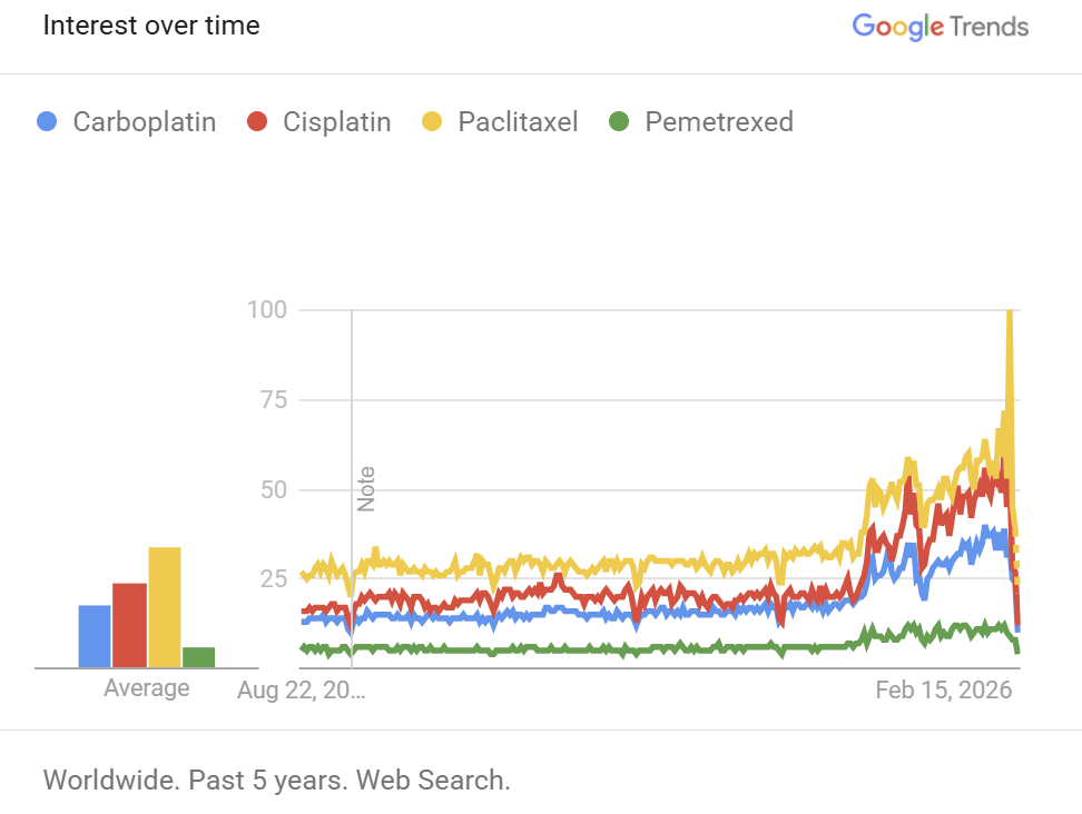
Chemotherapy APIs are the foundational cancer‑fighting drugs that work by damaging rapidly dividing cells, including tumor cells. Unlike targeted therapies, which act on specific mutations, chemotherapy affects a broad range of cellular processes. In lung cancer, platinum agents (carboplatin, cisplatin), taxanes (paclitaxel, docetaxel), and antifolates (pemetrexed) remain essential components of treatment. They are used across early‑stage, locally advanced, and metastatic disease, often forming the backbone of combination regimens with immunotherapy or targeted therapies.
How chemotherapy works
Chemotherapy drugs interfere with cell division and DNA repair. Platinum agents create DNA crosslinks that prevent cancer cells from replicating. Taxanes disrupt microtubules, freezing cells in mitosis and enhancing the effects of DNA‑damaging agents. Antifolates like pemetrexed block folate‑dependent enzymes needed for DNA synthesis. Because these mechanisms target fundamental cellular processes, chemotherapy can shrink tumors even when no actionable mutation is present — and it remains the standard treatment after targeted therapy resistance.
Evolution and role in modern treatment
Chemotherapy does not have “generations” like targeted therapies, but its role has evolved significantly. Historically used alone, it is now frequently combined with immunotherapy to boost immune activation, or paired with targeted therapies in specific molecular settings. Regimens such as carboplatin‑pemetrexed and carboplatin‑paclitaxel (“CarboTaxol”) are widely used and well‑studied. Even in the era of precision oncology, chemotherapy remains indispensable — providing reliable, broad‑acting treatment when targeted options are unavailable or no longer effective.
| API | Brand Name(s) | Target | Mechanism |
|---|---|---|---|
| Carboplatin | Generic | DNA | Cross‑linking |
| Cisplatin | Generic | DNA | DNA adduct formation |
| Paclitaxel | Taxol | Microtubules | Prevents cell division |
| Pemetrexed | Alimta | Folate pathway | Blocks DNA synthesis |
Chemotherapy Twitter (X) Insights
| Drug / Topic | Effectiveness Signals (easy to understand) | Side Effect / Concern Signals | Management / Community Insights |
|---|---|---|---|
| Carboplatin | – Backbone of many lung cancer regimens – Strong results when paired with pemetrexed or paclitaxel – Used as control arm in major trials (HARMONi, DESTINY‑Lung04) | – Hypersensitivity reactions occasionally mentioned – Fatigue and blood count drops appear in patient posts | – Oncologists explain why carboplatin is preferred over cisplatin in many lung cancer settings |
| Pemetrexed | – Key partner for carboplatin in non‑squamous NSCLC – Appears in multiple trial combinations (EGFR, HER2, MET) | – GI issues and fatigue mentioned indirectly | – Often referenced in sequencing after targeted therapy failure |
| Paclitaxel | – Strong synergy with carboplatin (“CarboTaxol”) – Used in lung, ovarian, and breast cancer regimens | – Neuropathy frequently mentioned – Bone pain appears in patient discussions | – Educational posts explain paclitaxel’s mechanism (cell‑cycle arrest → enhances platinum damage) |
| Cisplatin | – Standard in bladder cancer and chemoradiation regimens – Appears in historical lung cancer discussions | – Kidney toxicity and nausea mentioned indirectly | – Often discussed in “cisplatin‑eligible” vs “cisplatin‑ineligible” patient selection |
| Broader chemotherapy themes | – Chemo is the “backbone” against which targeted therapies are compared – Still essential after TKI failure | – Toxicity management (neuropathy, edema, GI issues) widely discussed | – Many posts explain why certain doublets (CarboTaxol, Carbo‑Pemetrexed) work well – Steady discussion volume in thoracic oncology (#LCSM, #NSCLC) |
Chemotherapy Reddit Insights
| API | Positive Themes | Negative Themes | Common Patient Discussions |
|---|---|---|---|
| Carboplatin | – Strong tumor shrinkage (large tumors reduced dramatically) – Manageable treatment compared to other chemo – Effective for recurrence prevention (testicular cancer) | – Nausea lasting days–weeks – Fatigue and brain fog – GI issues (diarrhea, loose stools) – Low blood counts requiring boosters | – Anti‑nausea meds taken proactively – Hydration + small meals – Comparing Carboplatin vs more aggressive regimens – Monitoring blood counts |
| Cisplatin | – Significant tumor shrinkage – Highly effective when combined with radiation – Strong benefit for younger patients | – Hearing loss + tinnitus – Severe nausea/vomiting – Fatigue, nerve pain – Kidney toxicity risk | – Aggressive hydration (IV fluids) – Adjusting anti‑nausea meds (e.g., olanzapine) – Reporting all symptoms early – Understanding cisplatin’s role as adjunct therapy |
| Paclitaxel | – Major tumor shrinkage (breast cancer foci disappearing) – Helps prevent recurrence – Effective across multiple cancers | – Neuropathy (hands/feet) – Fatigue + bone pain – Hair loss (head + eyebrows/lashes) | – Cold therapy during infusion to prevent neuropathy – Steroids masking early fatigue – Hydration + oral care to prevent mouth sores – Pain management (Claritin + Tylenol) |
| Pemetrexed | – Tumor shrinkage + remission – Rapid symptom improvement (cough, chest heaviness) – Standard of care with strong survival benefit | – Fatigue (persistent) – GI issues (constipation → diarrhea) – Mucus membrane irritation, dry mouth, eye burning | – Anti‑nausea meds around the clock – Hydration + protein intake – Easy‑to‑digest foods (yogurt, ice cream, juice) – Reporting symptoms proactively |